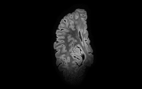
The right hemisphere and brainstem are scanned using ultra-high-field strength MR imaging (7 Tesla) in LUMC, Leiden.
The post-mortem 7T MRI (~0.5 mm3) will allow us to determine integrity of white matter bundles and grey matter nuclei in the brainstem and (sub)cortical regions at a resolution sufficient to distinguish separate nuclei and the tracts between them and define small grey or white matter lesions in the aging brain. T2-relaxometry, FLAIR, Susceptibility Weighted Imaging MRI (SWI), Magnetization Transfer Ratio (MTR) are performed, to quantify iron deposits and myelin/axonal changes.